Saving lives with technology, the MRI evolution on the healthcare
Nowadays, every field is deeply influenced and dependent on technology. The medical field is no exception. In fact, technology is fundamental to innovation and improvement in the diagnostic pathway: more accurate diagnoses contribute to more effective treatment pathways. One of the areas where technology has most revolutionized the medical field is MRI, where the introduction of new machines has resulted in significant breakthroughs.
MRI technology
Thanks to eXP reconstruction, physicians can analyze images in greater detail and obtain more precise diagnostic information, consequently improving patient care quality. Another key aspect of this innovative MRI technology is the MAR (Metal Artifact Reduction) feature, which facilitates imaging patients with metal implants, such as hip, knee or shoulder replacements. In the past, the presence of these metal implants could cause artefacts on images, making correct interpretation difficult and affecting the accuracy of diagnosis. The MAR function, on the other hand, has significantly reduced this drawback, allowing clinicians to obtain sharper and more detailed images, even in patients with metal implants.
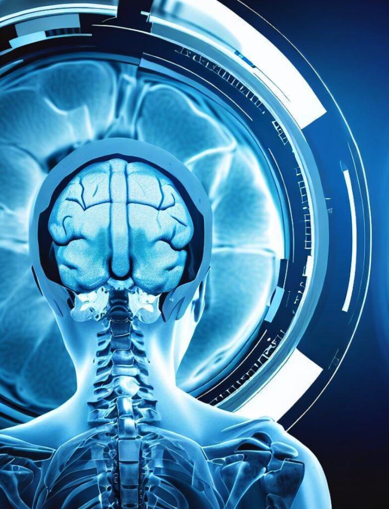
Starting with MRI, there are many other developments positively conveyed by technological innovation. For example, the so-called fMRI or functional magnetic resonance imaging. Functional magnetic resonance imaging (fMRI) is an advanced MRI technique that can help detect brain areas responsible for specific human functions, such as speech and language generation, motor function, movement, and vision.
What it is about and the benefits
This scan allows doctors to measure blood flow in the brain without surgery or using more invasive methodologies. fMRI imaging is a safe way to diagnose certain pathological conditions and determine whether certain therapeutic treatments work. A functional magnetic resonance imaging (fMRI) scanner detects brain activity using a powerful magnetic field. When an area of the brain becomes more active, such as when you shake your hand, there is an increase in blood flow to the region of interest. An fMRI imaging scan exploits active neurons that require more oxygen from red blood cells; this increase in activity leads to a change in blood flow detected by fMRI.

By indirectly measuring changes in blood flow and electrical activity, fMRI assesses a patient’s brain activity. Clinicians consider this type of indirect measurement a method to study blood-oxygen-level-dependent (BOLD) responses. Investigations with fMRI can study a wide range of cognitive processes, including decision-making and memory. The fMRI scans create a color-coded map of the patient’s brain activity. There are multiple advantages to performing this type of brain scan. It is a non-invasive procedure that does not require surgery, can detect abnormalities present in the brain, and helps to assess both the structure and function of the brain, allowing analysis of abnormalities hidden even behind bony structures.
The “feeling” scan
The fMRI scan is based on the same technology as MRI: both use a powerful magnetic field and radio waves to produce images of the body. However, there are important differences between the two; MRI captures images of the brain’s structure: it can see cysts, tumours, bleeding, and structural abnormalities. In contrast, fMRI acquires images of brain activity while performing a particular function. It turns out that it is even possible to analyze the patient’s reaction to certain thoughts and feelings. Functional MRI creates a functional map superimposed on images of the brain.
How it works
And then there is the technology used by Prenuvo, a brand that is very famous thanks to an Instagram post by Kim Kardashian. Prenuvo received financial backing from Cindy Crawford and her husband, Rande Gerber, who offers whole-body preventative magnetic resonance imaging, or MRI, exams without a physician’s order. Prenuvo claims that its precision MRI technology can detect over 500 conditions and that its scans result in a “lifesaving diagnosis” for one in every 20 patients.
Prenuvo uses proprietary software and artificial intelligence tools to make its whole-body scans different from other hospital scans. Prenuvo uses a type of MRI called diffusion-weighted imaging that measures tissue hardness, and cancerous tumours can have a higher tissue density. For some lung tumours, higher density can suggest growth is more cancerous. Although these imaging exams are legitimate tools utilized daily in the medical field, their use is often guided by a licensed medical professional. Companies like Prenuvo do not require that patients have a physician’s order to complete one of their scans. If you can pay, you can play.


